
Bone density: The “barometer” of bone strength
Bone Mineral Density (BMD) is a core indicator for measuring bone strength, assessing the degree of osteoporosis and predicting the risk of fractures. Bones are not static but are in a dynamic process of “reconstruction” : osteoblasts are responsible for synthesizing new bone, while osteoclasts break down old bone. When young, osteoblasts are active and bone mass gradually accumulates, reaching its peak around the age of 30. After that, the rate of bone loss gradually exceeded the rate of formation, and bone density began to decline. If this process is out of balance, it may cause osteoporosis.

Osteoporosis: The Silent “Invisible Killer”
Osteoporosis is characterized by reduced bone mass and destruction of bone microstructure, and is known as the “silent disease”. Because it has no obvious symptoms in the early stage, it is often discovered only after a fracture. Its harm should not be underestimated:
1. sharp increase in the risk of fractures: The “brittle collapse” of bones
The most direct harm of osteoporosis is a significant increase in the risk of fractures. Normal bones are like dense honeycomb structures, while osteoporotic bones are like wooden boards eaten by insects. Trabeculae become thinner, broken, and have more pores, resulting in a decrease in bone strength.
Common fracture sites:
Hip fracture (femoral neck or intertrochanteric) : Known as “the last fracture in life”, the mortality rate of patients within one year is as high as 20%, and more than 50% will permanently lose the ability to walk independently.
Spinal compression fractures: The vertebral bodies are like flattened biscuits, which may cause severe back pain, height reduction (up to 3 to 6 centimeters), and hunchback (” rickets “).
Wrist (distal radius) fracture: It is prone to occur when using hands to support the ground during a fall, affecting daily grasping and movement.
2. Posture deformities and Functional Disorders: “Irreversible Damage” to the Body
Vertebral compression fractures caused by osteoporosis will gradually change posture and physiological functions:
Kyphosis (kyphosis of the spine) : Excessive curvature of the thoracic vertebrae compresses the chest cavity, leading to a decrease in lung capacity and shortness of breath. In severe cases, it may even affect heart function.
Abdominal compression: Deformation of the lumbar vertebrae squeezing the abdominal cavity may cause abdominal distension, constipation and loss of appetite.
Chronic pain: Persistent pain caused by fractures or bone deformities, which intensifies with activity and reduces the quality of life.
High-risk groups: The interweaving of age and risk
Osteoporosis is not exclusive to the elderly, but the risk is significantly increased in the following groups of people:
1.Postmenopausal women: Estrogen has a protective effect on bones. After menopause (around the age of 50), estrogen levels drop sharply and bone loss accelerates. Data shows that the prevalence of osteoporosis among women over 50 is 32.1%, and the risk of fractures is four times that of men.
2. Men over 70 years old: The slow decline of androgen leads to a gradual decrease in bone density, and the incidence rate rises after the age of 70. Osteoporosis in men is more insidious and often leads to delayed treatment due to the neglect of screening.
3. Young high-risk groups: People who have been using glucocorticoids for a long time, lack exercise, smoke, drink excessively, have vitamin D deficiency, or suffer from hyperthyroidism, diabetes and other diseases may experience bone mass loss earlier. Recent studies have found that vitamin D deficiency caused by prolonged sitting and excessive sun protection leads to bone density in some young people being lower than that of their peers.
Bone density testing: Gold standard
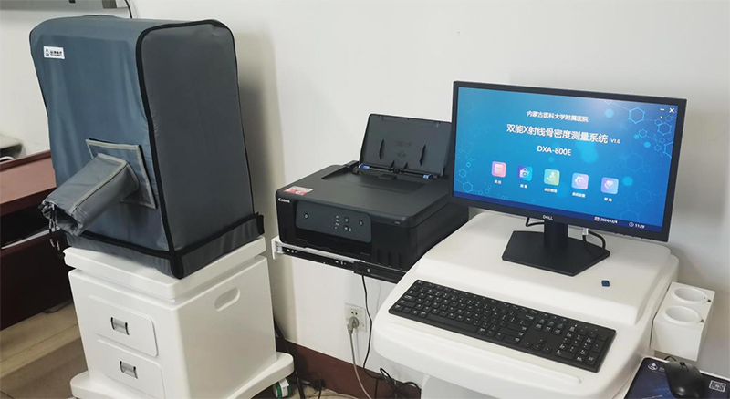
Early diagnosis is the key to preventing and treating osteoporosis, and Dual-energy X-ray Absorptiometry (DXA) is internationally recognized as the “gold standard” for bone mineral density testing.
The principle is to calculate the mineral content by penetrating the bones with X-rays of two different energies. DXA has the advantages of low radiation, high precision and quick operation, and can accurately measure the bone density of the lumbar vertebrae and hip. When diagnosing bone mineral density values, the “T value” is used as the evaluation criterion: The bone mineral density of the patient is compared with the peak bone mineral density of healthy young people. A T value ≥-1 is considered normal, -1 to -2.5 indicates decreased bone mass, and ≤-2.5 is diagnosed as osteoporosis.
Post time: Apr-28-2025