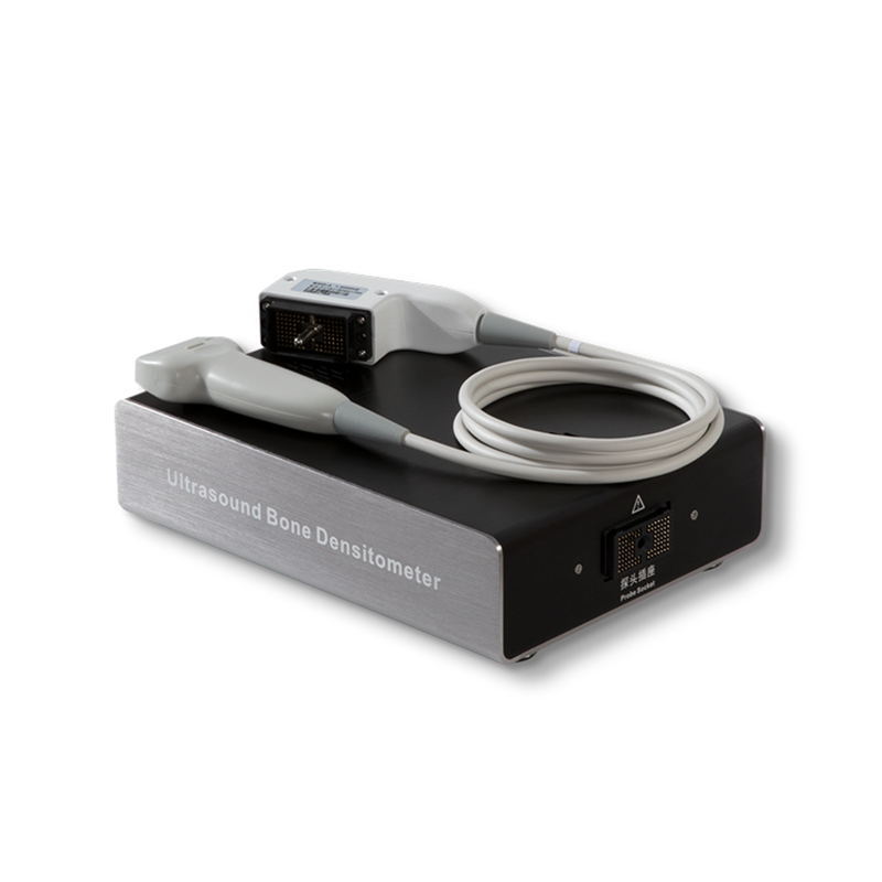
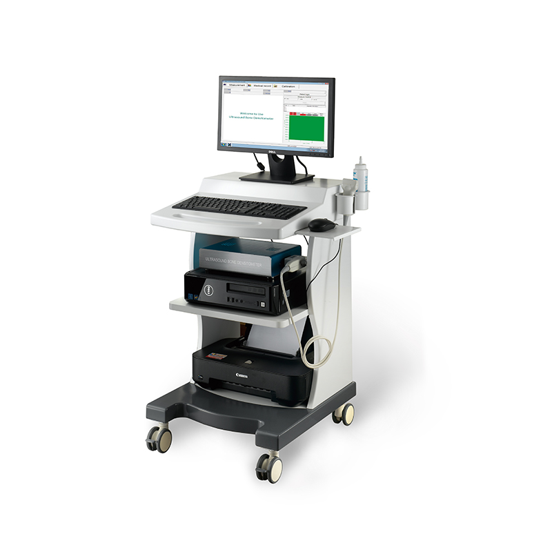
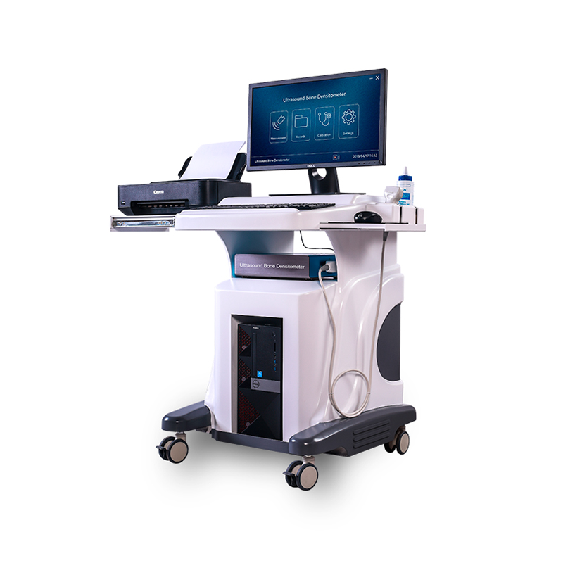
2022 Good Quality Bone Densitometry Other Term - Dual-Energy X-ray Absorptiometry Bone Densitometry DXA 800F – Pinyuan
2022 Good Quality Bone Densitometry Other Term - Dual-Energy X-ray Absorptiometry Bone Densitometry DXA 800F – Pinyuan Detail:
Application
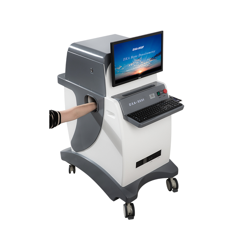
A bone density test is used to measure bone mineral content and density. It may be done using X-rays, dual-energy X-ray absorptiometry (DEXA or DXA), or Ultrasound to determine bone density of the radius, tibia and forearm. For various reasons, the DEXA scan is considered the “gold standard” or most accurate test.
This measurement tells the healthcare provider whether there is decreased bone mass. This is a condition in which bones are more brittle and prone to break or fracture easily.
Features
Large Scale Integrated Circuit
Multi -Layer Circuit Board Design
Light Source Technology With High Frequency and Small Focus
Imported High Sensitivity Digital Camera
Using the Cone – Beam and Surface Imaging Technology
Using Laser Beam Positioning Technique
Using the Unique Algorithms.
ABS Mould Manufactured, Beautiful , Strong and Practical
Special Analysis System Based on Different Countries People
Technical Specifications
Using digital Laser Beam Positioning Technique
Special Analysis System Based on Different Countries People
Using the Most Advanced Cone – Beam and Surface Imaging Technology.
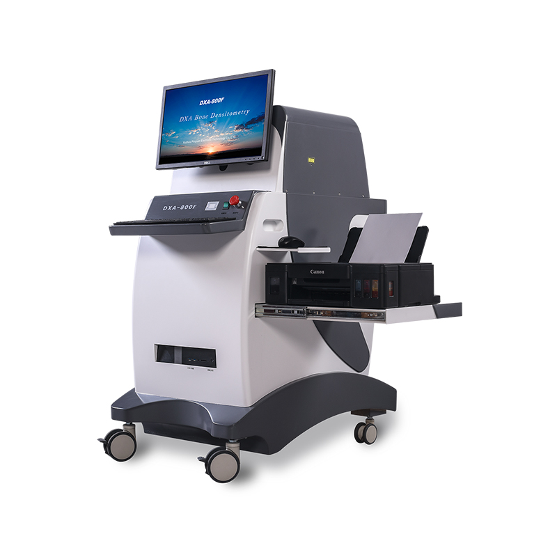
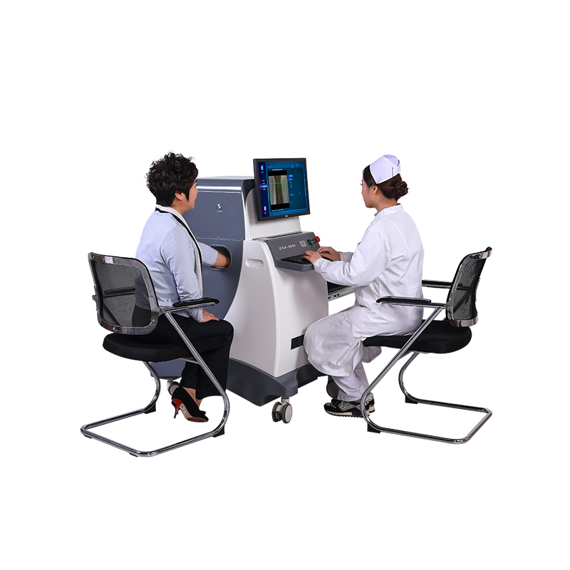
Measurement Parts: the Front of Forearm
With High Measurement Speed and Short Measurement Time.
Adopting the Full Closed Lead Protective Window to Measure
Technical Parameter
1.Using the Dual Energy X-ray Absorptimetry.
2.Using the Most Advanced Cone – Beam and Surface Imaging Technology.
3.With High Measurement Speed and Short Measurement Time.
4.With Dual Imaging Technology to Get More Accurate Measurement.
5.Using Laser Beam Positioning Technique, Making the Measuring Position More Accurate.
6.Dectcing Image Digitization, to Get Accurate Measurement Results.
7.Adopting the Surface Imaging Technology, Measuring Faster and Better.
8.Using the Unique Algorithms to Get More Accurate Measurement Results.
9.Adopting the Full Closed Lead Protective Window to Measure, only Need to Put the Patient’s Arm into the Window. The Equipment is Indirect Contact with the Scanning Parts of the Patient. Easy to Operate for the Doctor. It is Safety for the Patient and Doctor.
10.Adopting Integrated Structure Design
11.Unique Shape, Beautiful Appearance and Easy to Use.
Performance Parameter
1.Measurement Parts: the Front of Forearm.
2. X ray tube voltage:High Energy 70 Kv, Low Energy 45Kv.
3.The high and low energy corresponds to the current, 0.25 mA at high energy and 0.45mA at low energy
4.X-Ray Detector:Imported High Sensitivity Digital Camera.
5.X-Ray Source:Stationary Anode X-ray Tube (with High Frequency and Small Focus)
6.Imaging Way:Cone – Beam and Surface Imaging Technology.
7.Imaging Time:≤ 4 Seconds.
8.Accuracy(error)≤ 0.40%
9.Repeatability Coefficient of Variation CV≤0.25%
10.Measuring Area :≧150mm*110mm
11.Can be connected to hospital HIS system, PACS system
12.Provide Worklist Port with independent upload and download function
13.Measuring Parameter: T- Score, Z-Score, BMD、BMC、 Area,Adult percent[%], Age percent[%], BQI (The Bone Quality Index) ,BMI、RRF: Relative Fracture Risk
14. It with multi race clinical database, including: European, American, Asian, Chinese, WHO international compatibility. It measuring the people between the age of 0 and 130.
15.Measuring children over three years older
16.Original Dell Business Computer: Intel i5,Quad Core Processor, 8G, 1T, 22’inch HD Monitor
17.Operation System: Win7 32-bit / 64 bit, Win10 64 bit compatible
18.Working Voltage: 220V±10%, 50Hz.
Why Might I Need A Bone Density Test?
A bone density test is mainly done to look for osteoporosis (thin, weak bones) and osteopenia (decreased bone mass) so that these problems can be treated as soon as possible. Early treatment helps to prevent bone fractures. The complications of broken bones related to osteoporosis are often severe, particularly in the elderly. The earlier osteoporosis can be diagnosed, the sooner treatment can be started to improve the condition and/or keep it from getting worse.
A bone density testing may be used to:
Confirm a diagnosis of osteoporosis if you have already had a bone fracture
Predict your chances of fracturing a bone in the future
Determine your rate of bone loss
See if treatment is working
There are many risk factors for osteoporosis and indications for densitometry testing. Some common risk factors for osteoporosis include:
Post-menopausal women not taking estrogen
Advancing age, women over 65 and men over 70
Smoking
Family history of hip fracture
Using steroids long-term or certain other medicines
Certain diseases, including rheumatoid arthritis, type 1 diabetes mellitus, liver disease, kidney disease, hyperthyroidism, or hyperparathyroidism
Excessive alcohol consumption
Low BMI (body mass index)
What Are The Benefits Vs. Risks?
Benefits
● DXA bone densitometry is a simple, quick and noninvasive procedure.
● No anesthesia is required.
● The amount of radiation used is extremely small—less than one-tenth the dose of a standard chest x-ray, and less than a day’s exposure to natural radiation.
● DXA bone density testing is currently the best standardized method available to diagnose osteoporosis and is also considered an accurate estimator of fracture risk.
● DXA is used to make a decision whether treatment is required and it can be used to monitor the effects of the treatment.
● DXA equipment is widely available making DXA bone densitometry testing convenient for patients and physicians alike.
● No radiation stays in your body after an x-ray exam.
● X-rays usually have no side effects in the typical diagnostic range for this exam.
Risks
● There is always a slight chance of cancer from excessive exposure to radiation. However, given the small amount of radiation used in medical imaging, the benefit of an accurate diagnosis far outweighs the associated risk.
● Women should always tell their doctor and x-ray technologist if they are pregnant. See the Safety in X-ray, Interventional Radiology and Nuclear Medicine Procedures page for more information about pregnancy and x-rays.
● The radiation dose for this procedure varies. See the Radiation Dose in X-Ray and CT Exams page for more information about radiation dose.
● No complications are expected with the DXA procedure.
Product detail pictures:

Related Product Guide:
We are ready to share our knowledge of marketing worldwide and recommend you suitable products at most competitive prices. So Profi Tools offer you best value of money and we are ready to develop together with 2022 Good Quality Bone Densitometry Other Term - Dual-Energy X-ray Absorptiometry Bone Densitometry DXA 800F – Pinyuan , The product will supply to all over the world, such as: Romania, Naples, Japan, We integrate all our advantages to continuously innovate, improve and optimize our industrial structure and product performance. We will always believe in and work on it. Welcome to join us to promote green light, together we will make a better Future!
We have been cooperated with this company for many years, the company always ensure timely delivery ,good quality and correct number, we are good partners.

